Hello, it's Anthony Chadwick from the webinar vets, welcoming you to another one of our webinars. One of our value words at the webinar best is innovation, and the company that I want to introduce you today to today is, has this in bucket loads. Very fortunate to meet them at the London Vet Show, and their product has really just blown me away.
And I'm really fascinated to see how everybody else finds it later on, during the, the webinar. This is really about point of care ultrasound. There's no magic in this, there's no hocus pocus, there is just pocus.
So it's point of care ultrasound. And we're going to hear from Joanna and Katie a little bit later, but first of all, we've got a video to show you, which is presented by Doctor Richard Markle and Doctor, Doctor Robert Fulton. Richard is a 1985 veterinary graduate from Missouri University, and he's the CEO of Aluminix X Consulting.
He works with the US Olympic equestrian veterinary team, with the, sorry, the US Olympic equestrian team as one of the vets. And he's a frequent speaker at the American Association of Equine Practitioners Congress. He lives in California at the beach with his wife, son, and one naughty dog and one nice dog.
Robert is a University of Georgia alumni, also a vet. He specialised in his career in emergency and critical care medicine and also reproduction. He ran an outpatient ultrasound department at a at a referral hospital in Virginia and has written several chapters in focused ultrasound techniques for small animal practitioners and point of care ultrasound techniques.
He's passionate about teaching ultrasound. I think this whole concept of point of care ultrasound can be really revolutionary in, The veterinary field, and I'll be really interested to hear what everybody else thinks as well. So, looking forward to speaking to everybody later in the question and answers, but we're now going to run a video with Richard and Robert, who will explain the, how this works better.
So, over to the video. Welcome and good afternoon everyone. At Butterfly, we are passionate about our mission to democratise imaging around the world for all health care practitioners by providing valuable clinical information that improves decision making at the time and place it is needed.
This mission is one that we take very seriously as our team obsesses over ways that we can make our products and services better and more accessible for all clinicians and patients around the world. With this said, today we have very special news to share with our partners and customers in the veterinary community. Almost 2 years ago we introduced our innovative veterinary product, the IQ vet, to the US market.
With IQVett we began to create a positive shift in veterinary care, highlighted by examples like Texas Tech University's decision to empower every incoming veterinary student with an IQ vet. And our recent partnership with MWI Animal Health to expand our commercial reach in the US. In the last year, we've also made significant investments within our company to build a fully dedicated and deeply passionate expert veterinary team focused on supporting innovation in animal health care with butterfly technology.
While the veterinary market represents an exciting opportunity for butterfly to continue to innovate, it's also very personal to me as an owner of both dogs and horses. In equestrian sport, I've experienced firsthand the importance of point of care information in clinical decision making and the benefit of making quicker and more informed decisions in situations where a traditional ultrasound system simply cannot go. So I am very excited about what butterfly can do today and in the future for animal healthcare and the practise of veterinary medicine.
Butterfly IQ vet is already proving to be a highly useful tool because when patients cannot speak, an image is worth 1000 words. Butterfly makes point of care ultrasound or pocus practical by allowing practitioners to incorporate valuable ultrasound information into routine clinical decision making because seeing is knowing and butterfly makes it possible. What is even more exciting beyond butterfly's value as an advanced assessment tool is its ability to change the way clinical interactions occur.
Butterfly allows veterinarians to connect with owners by showing them what the issues are, making it easier to understand what is going on and moving from the theoretical discussion to visual explanation. Ultimately with butterfly, veterinarians like you can better understand your patients and bring the technology anywhere you need to go, wherever your patience may be. With this foundation, I am very excited to share a big advance in the ability for veterinarians to see and know better than ever before.
Today we are announcing the global launch of a revolutionary technology and the next big shift in veterinary medicine. The butterfly IQ plus vet. No.
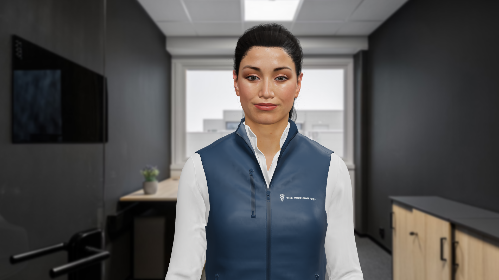
Yeah. Hi, I'm Jenna Mutch. I'm the senior director and head of commercial for the veterinary business of Butterfly.
Today, Katie Moore joins me, our clinical product specialist for our veterinary business. For over 50 years, ultrasound machines have used fragile and expensive piezo crystals, each wired and attached with cables to a separate machine. Those crystals operate at a limited frequency band and require different transducers that allow veterinarians to capture specific imaging depths to view different parts of the body on different species.
These traditional cumbersome systems can cost upwards of $50,000. Butterfly has reinvented ultrasound technology from the ground up. Replacing these crystals with a single silicon chip, what we call our ultrasound on chip technology.
The chip, made from an array of thousands of programmable micromachine sensors. You can think of this as a grid of very small drums, is capable of emulating all three ultrasound wave patterns with a single probe, all in one handheld device. Through this innovation, Butterfly is able to drastically lower the cost of the device by leveraging the same manufacturing consumer products, allowing for high precise, high quality ultrasound to be produced at scale, making medical imaging more accessible and affordable than ever before.
I'm so excited about our IQ plus vet and Katie. Doctor Fulton and Doctor Markel are here to tell you why. Thank you, Jenna.
I'm very excited to be here tonight to talk to you further on the ultrasound on chip technology and what it brings to the advancements of the IQ plus that. Starting with our abdominal preset, we are able to actually increase our field of view, not only allowing you further assessment of each abdominal organ, but give you the most information that you need at first glance. Our abdominal preset deep is also allowing us to have actually better penetration up to 30 centimetres along with also enhancing our resolution in both far and near fields.
We are also able to improve our Doppler sensitivity to aid you in vascular assessments, but also now give you an added bonus to be able to detect ureteral jets like seen in this clip into the bladder, which is your first clinical suspicion that if you lack this jet, you could have an issue like an atopic ureter. Moving to our equine presets and muscle skeletal presets, we also did image optimisation to work on both tendon and ligament delineation along with muscle fibre patterns. We too also enhanced our bone interface feature to help with procedural guidance to find those bony landmarks.
And now, I am very excited to say that we are compatible on on select Android devices, which brings us back to our founder's first statement of wanting to provide medical imaging to all. Our smaller probe and smaller probe size and footprint in size as well helps guide our patients a little bit more comfortably through the ultrasound exam. And in doing so, though, we made our probe just slightly smaller, we actually were still able to increase our battery life and therefore also increase our continual run scan time.
And we know in the vet field we're tough on our equipment, so we made sure that we have a probe that keeps up with us. We have a 4 4 ft drop durability, along with water resistance and dust resistance as well. I am also very excited to say that our needle guidance tool for in-plane use is called Needlevis.
This helps you not only for novice scanners, when first trying to get to that region of interest, highlight that needle to make it more visible, but also for those more efficient, be able to visualise it in superficial areas. And my best and most loved feature of this, and I think which makes us most unique to Butterfly is our tele guidance, allowing you, the practitioner, to not only be able to sit remote from your office or from your home and guide your colleague in the field or your technician. So, at home, you will be able to see on one side of your screen, the ultrasound image, and on the other side of your screen, be able to actually see where that transducer is placed on your patient.
You too at home will be able to adjust the image, including gain, depth, and notation and image archive. And I'm very excited to now introduce Doctor Robert Fulton, who is a small animal practitioner with a focus on pocus and zoo medicine. Hi, I'm Robert Fulton.
I've been using ultrasound in one degree or another since 1994 from early early parts of my career with equine and small animal reproductive scans into almost 18 years in the emergency and critical care arena where ultrasound is such a useful tool, the past 10 years in a progressive small animal practise. I've also been teaching ultrasound since about 2001. And so I've seen ultrasound over my career progress not only in the technology that we have but also in how we use ultrasound.
For about eight years now, it's been specifically teaching Focus of point of care ultrasound. And that is really starting to expand how ultrasound can be used in our, in our clinical practise, to the betterment of our medicine, of our patients, and the, and our clients as well. So let's start out with a little video clip I think we have of a of a gallbladder.
So here, this is obtained with the butterfly IQ plus VAT. And this was in a 13 year old female spayed miniature schnauzer presented for lethargy, some vomiting, and hypoorexia. As part of our workup, here's the ultrasound, and we can see in this image a really nice view of the gallbladder is what we're looking at.
There we're seeing a little bit of the common bile duct which looks rather distended. This gallbladder is pretty round and filled with this. Genic sludge.
We can see lots of detail in that wall. It's still nice and thin and smooth. And this is information that we can obtain right here in the, in the exam room.
I do appreciate your point that you say that you can not only detect this in the exam room, but knowing that at least when we talk about this type of finding of that consolidation to the gallbladder. It's a very significant thing to be able to actually detect this early because we know as this progresses, our outcome diminishes quite completely. So I, I do totally appreciate about that detection that you can have early on for this type of condition.
And so here this is a great example of how ultrasound in general can forward our patient care, and we're able to forward that even faster because we can do this truly point of care. As I've been teaching point of care over the past several years, some of the catchphrases are modern day stethoscope or an extension of the physical examination. But still, since most veterinary practitioners have console machines or even laptop sound machines, it is more the case that we still take the patient from the exam room to the ultrasound equipment with handheld units like the butterfly.
Now we're Truly taking the the modality to the, to the point of care to the exam room. And so it really opens up the, the point of care, the way that we are that we think about the point of care as well. With these, with these scans and doing that in the exam room with a nice portable machine like the butterfly, we can show the client these images while we're obtaining them so we can engage the client, we can increase the value to our client in addition to forwarding our.
Or information about the about our patient. So where I used to teach point of care as thinking about as part of our minimum database, we'll do a physical examination and then we'll do CBCIM UA and Pocus. Now we are truly with butterfly.
Take it into take that step forward to making an extension of the physical examination exactly the same way that we use a stethoscope to extend our sense of hearing into the patient. Now we can extend our sense of sight into our patient right there in the exam room. I completely agree, and I, I totally am in want to also one, love that minimum database that you can collect pretty early, so you can, again, further your, your diagnostics from there and actually help guide.
And now to introduce Doctor Richard Markel, who is an equine practitioner. Hi Katie, thank you so much for having me and, and the opportunity to, to talk about point of care ultrasound and ultrasound, . I'm a veterinarian.
I focus in sports medicine for horses. I've been one of the veterinarians for the US Olympic team for quite a few years, as well as, I think I've done 7 World Cup show jumping andresage finals now is either treating your team that and Ultrasound is a staple in, in my, in my practise. I mean, I couldn't imagine going to work and seeing my patients without a, as Doctor Fulton said, an ultra or a stethoscope and, and a thermometer and, and my 300 years of experience.
But, now I can't imagine doing it without, without an ultrasound and a butterfly, and it's really been an evolutionary shift, diagnostic. Being so portable and being affordable and, and having a horse kick it or drop it, and it's tough as nails. And I would say I've at least tripled the number of scans that I do because if I feel a lump or bump, it's really easy for me to, not take a look at it.
I think on top of that, I, I can't imagine. Putting a needle for an injection, of a joint or an abscess or a biopsy without an ultrasound guide. So it really changed mechanically how I approach every case.
So, before we look at a case, I just wanna mention to you, you know, ultrasound isn't easy. I've been doing it for a lot of, a lot of years and it's still tough sometimes, but what the engineers at Butterfly have been able to do and, and Katie, with your help and the engineers and the doctors that have looked at images. They've been able to kind of have the equivalent of a point and shoot machine.
And I'm, I'm kind of excited about that because the images I'm gonna show you today, literally alcohol, ultrasound gel, and reset the probo. So with those presets, it makes it quite easy. If you're an advanced user, we certainly have tools in the software that you can change and optimise your image, but the idea was to be able to look at things quickly and easily and have a really good diagnostic image.
So, I'm ready to jump into those scans. Perfect. So this is a, this is a 12 year old Grand Prix show jumper, and, and Katie, I don't do a lot of video imaging except when I'm saying a pre-purchase exam or I'm doing a quick scan.
Most of them, I'm focusing on a particular scan and want to look at something. But let's go through this video here and as we just scanned down the leg and I think we all think about ultrasound as a soft tissue imaging device. And, and, and I always try to teach when I am teaching ultrasound, is we need to look at that bone because ultrasound is a wonderful bone imaging tool and oftentimes can see abnormalities.
For example, the The holy grail of, of headaches and, and our sports medicine equine practise lives is that pros dispensary ligament. And it, we see the injuries to that bone insertion as we do to the ligaments. And there are sometimes, coexisting morbidities.
So, you want to see that really nice sharp bone, that very clear line that's just above my, my annotation there, as well as, of course, looking at your fibre alignment, your fibre pattern, your echo density, and fluid that we can see that normal, fluid underneath that deep flexor tendon there. So, these are just, this is, probably a 1250 pound horse, And like I said, middle-aged and, and we just put the, the gel and alcohol on there. That little bit of feathering that you'll see right at the top of your screen, that little feathering is just an artefact because we're using a standoff pad here.
So when I'm doing my distal limbs, I will use a standoff pad, when I'm doing cervical. Facets or sacroiliac injections, other deep injections. If I'm scanning a chest or, or heart or lungs, or doing an abdominal fat scan, we don't use, we don't use the standoff pad for those distal limbs, it's really nice because the, the image has been optimised with that.
So, great image, I've, I have used literally some of the most advanced expensive machines over the years since 1987, . 88, 89, and it's just remarkable, the evolution in technology and I don't want to sound overly dramatic, but, you know, this is, this portability is as revolutionary for us as digital radiographs were and doing MR and CT technology. So it's really exciting to be able to have this in, in my, in just right in my hand and a two-second boot up and I'm ready to go.
I completely agree. Being able to see you work in the field, I 100% appreciate now how much that portability really is necessary. And since you're, since you focus on small animals, you can see it's also important for us to be able to get out of the way quickly.
So these are, these are just a couple of images and, and gosh, I, I wanted an hour today and they told me I couldn't have an hour, but there's so many images and cases that I, I just really want to talk to you about. And we're gonna be putting some on the website over time so we build some education behind this and, and really give veterinarians a little bit of a, a leg up on, on understanding and using ultrasound. And reference images.
But on the left, we have, again, point and shoot. This is a superficial digital flexor tendon and a deep and very proximal. And what I wanted to show was not only the clarity and definition in the margins of the ligament, fluid echo, but on the right, you can see, I, I've almost gone completely.
Instead of doing circumferential areas, I'll do a cross-sectional area because the circumferential measurements are sometimes challenging to do. They're really challenging in the field, but, and, and actually this particular ligament, I, I believe, Katie, I, I sent to you right off the iPhone while I was doing it and, and that's, that's a really cool. That we can do, we can share with a colleague and say, what do you think of this?
We can send it to the, the client's phone right while we're standing there so they can have a copy of it or to one of your, your colleagues for another opinion. So great features and, and really easy to use. .
I think Katie, at the end of the day, you know, we have to talk about how, what what does butterfly do for my practise and how does it make a difference? And I really believe that using ultrasound regularly in your practise, having access to it so easily, will truly change you as a doctor. You become a better doctor and you have better patient outcomes.
And that's also why I'm so excited about being able to have such easy access to ultrasound. I agree. I obviously am very passionate about ultrasound, but from emergency medicine to just day to day use, I agree.
You, you definitely add more to your exam and more to your outcome if you can actually figure out more from that initial start and presentation. Well, thank you so much for having me and, and sharing that excitement that I have. I, I love to talk to, to veterinarians about it and, and people that really see how important ultrasound is for us taking care of those patients that we spend our lives trying to do a better job every day.
Well, thank you so much for taking the time and being here tonight and thank you also Doctor Fulton for being here as well. I so appreciate and love talking to you both. Thank you, Katie.
I'm thrilled to announce our keynote speaker for the next big shift, Dr. John Daciano. Hopefully I didn't mess that up.
But he is the dean of the Academic and Student Affairs at Texas Tech University. Super happy to be here. And, one of the things that, we have at Texas Tech.
Is, we're a new school, but we have two facilities. One's called Amarillo Campus and the other one's called Mariposa Station. And I don't even know if Katie, or Darby or Jenna know this, but Mariposa means butterfly in Spanish.
And so, we actually have a connection there. The next big shift. So for me, the next big shift was To go from, I was at Virginia Tech for most of my career, but now I'm at Texas Tech, so I'm at the other tech.
So I need a big shift in moving to different universities, but, it's to come here to Texas Tech to make a difference in student education. That's kind of what we're gonna talk about today and how ultrasounds integral to that part. This is my wife, Linda, next to me.
I've got Rader Red and the master writer there. So, super happy to be here at Texas Tech. Next slide, please.
So I was going to reminisce, but it seems like we all have done that already. This was a resident that I was instructing when I was at Virginia Tech, and we were doing a transabdominal ultrasound on this mayor who had a late-term gestation foetus, and we were doing foetal well-being assessment. And that ultrasound machine is, you know, probably close to $100,000 at the time.
We wanted it because we could do Doppler and some other aspects, but you can imagine, you know, just trying to get that away from the horse, if something were to go awry. So, you know, as, as the other speakers have said, ultrasound has come a tremendous way as far as technology to enable us to do all the things. That we used to be able to do with these massive machines we can now do just with a handheld.
So where are we with student education? So one of the big changes that happened in student education over the last 10 years is a shift to more clinical and professional skills, so more hands-on. We were very good at transferring knowledge to students, but we weren't so good maybe at transferring skills-based activities to them.
And practitioners would commonly say to us that And students would come out of school and I still needed to train them, in order to, to really be productive in my practise. And then students were saying that they needed to do internships because they didn't feel comfortable going into practise. So about 10 years ago, there was a great big shift in actually including more hands-on, physical activity, in our programmes and When you're able to build a new school like we are here at Texas Tech, we can include this purpose-built facilities to really enhance student education.
So, that training starts first semesters. This is just a picture of our first semester students actually learning surgery skills. And so the idea is that, you take those 1st 6 semesters, the pre-clinical education and you build upon their skills and allow them lots of time to practise.
So that by the time they get out into their clinical year, they're not, they're not actually naive about performing these activities. They're actually good at them and they can be productive members of a practise. And for us, our model is a little bit different.
We have a clinical year where the students are out amongst veterinary practitioners, and so, they need to be productive and need to be independent at that point. So, with, with all skills, including ultrasound, we're designing them over a 6 semester period to where it's almost like they're at the end of their, their senior year at the end of 3rd year. We want them to be very good at ultrasound.
We want them to be able to recognise all normals. We want them to be able to recognise some common abnormalities as well, and be able to drive the probe like it's nobody's business. So, there's some literature out there, about transferring skills to students and how you go about doing it.
There is no sharp improvement from one practise to the next. This is actually a casserole dish that I made. I like doing pottery, and it's taken me a long time to be able to make something like that.
I've destroyed probably more clay. That I've made, in order to create something like that. But it takes you a long time.
In music, in, science, in the arts, people will tell you it takes years of practise and 10,000 hours to become an expert at something. Certainly, you can be good at something, but you're probably a lifelong learner, with all of your skills that you have. Really the biggest improvements, that you see are associated with the quality and quantity of practise.
So, you can, you can start out with some natural talent. I don't know what that would be in an ultrasound is probably hand-eye coordination from playing video games, but really you're gonna, you're going to surpass that with deliberate practise. So we can take a student who is not so good at something and really make them very good at it with deliberate practise.
So, there's kind of three things that we do with skill acquisition. One of them is that what we've mentioned already is repeated performance. So that's why we start first semester.
We actually put ultrasounds in the hands of first semester students. So we want them to repeat it multiple times within a. And their cross-semester.
And then this rigorous skills assessment means that we have instructors who are good at ultrasound. Myself, I'm, was, mostly equine-oriented, so I did a lot of equine ultrasounds across my career. And you really need somebody who can guide those students to let them know, you know, where to go next, what they're looking at, How to get a better view.
And then you need what we call specific informative feedback. So we don't say, you know, well, that's great. You got it.
We would say, that's great, you got it, because, you know, you see this in your field, you can see that. So you get a very specific feedback so that way the next time they perform it, they're actually going to key in on that specific feedback to look for those things again. So we took the next logical step for us.
We purchased 55 butterfly ultrasounds. That's quite a, quite a number. I could never have done that 1015 years ago.
I just wouldn't have the budget to be able to do that. And so, our students all have iPads. We now know that, you can work with some, Android phones as well, but most students have some kind of device, but we required them to have an iPad when they came in.
And that was for a number of reasons, which also included the fact that we were purchasing, butterfly ultrasounds. We assign one probe to 2 students and they actually keep it. So it's in their possession.
They can practise on their own animals if they want to. And then we design and are creating, a curriculum where over time we're going to build those skills of those students. So, you know, when you are thinking about, you know, it's hard to always remember back when you learned something, but You know, many of us know that one of the big first steps is just being able to put the probe on an animal and actually find what you're looking for, let alone being able to assess it.
And so, again, having those, those probes in the hands of the students, but then also having them in the hands of faculty and staff, really helps us because then everyone's very familiar with the equipment and can, can help each other. We have a mission here at our school of serving rural and regional communities. Regional communities to us are small cities, areas away from large metropolitan areas which are often away from referral centres.
And so we're really trying to give those students the skills they need to be fairly independent. The other aspect of our programme is what's called, access and affordability, which was resonated earlier, in discussions about the butterfly probe as well. For us, access to education and affordability of education.
And so that also comes with a price point that we can afford an ultrasound to be able to put in the hands of students. And you know, it's, it's going to be important for us to be able to give those skills to the students where they can then be able to assess patients. On their own in, in practises and hopefully contribute to those practises.
So, as part of our distributed model of education, we have students in their clinical year out amongst practitioners. This is actually a picture of the butterfly team when they came and were assisting students to initial setup and to get them oriented to the machines. But we're, we're sending students out in our clinical year out amongst veterinarians.
And so they're working in these private practises. And what that allows us to do then is to get feedback from those practitioners on what is working well, what's not working well. It may be that the have more skill level at that point in ultrasound than what some of the practitioners may have.
How can we help them, the practitioners to become better at ultrasound, so they can assist the students? How can the students train some of those veterinarians? What is it that, we're missing in our educational model that can then enhance that experience?
And the skills for the students. So we'll, we'll seek that input. We'll have a very nimble curriculum that we can adjust so that we can continue to refine that, that education.
So expectations of our programme. We, we are expecting to partner with like-minded groups such as Butterfly. We hope to make ultrasound part of all examinations that we're doing, our point of care ultrasound, as we mentioned earlier.
We want our students to be those productive members and we want them to contribute, not just be, you know, someone who's sitting back watching, cases and, you know, people don't have confidence in their ability. We don't want that. We want them to be able to be contributing members and be able to help the practises that they go into.
And, you know, we'll have to figure out what happens when we do, exceed the training of some of our clinical partners and how we can help them to, to get to a higher level. And so that may be training we have with our practitioners separate from when we have students go there. So I just wanted to thank everyone for the opportunity to, to give a little glimpse into what we're doing with ultrasound training, how we're integrating butterfly into our programme.
As people have mentioned before, the excitement that everyone has about the ability to use some of these products that have really revolutionised, the ability to, to be mobile, to put them in situations. You know, you can put them in your pocket now and carry them around. So it's not like we're gonna have to run back to the clinic and see who has the ultrasound machine, and then we've got to put it in our truck and, and run somewhere with it, or, you know, we've got to wheel it down the hallway somewhere.
I mean, it's right there with us and makes it so much more accessible and thus, we're going to use it a whole bunch more. So, thank you so much for your time and all the best. Thank you so much, John.
Thank you for your vision. Thank you for your trust in us, and, and also thank you for taking care of your passion for the veterinary industry and taking care of our next generation. So we really appreciate it.
Up next, I'd like to introduce, to close us out, Darius Shahida. He's our chief strategy officer and chief business development officer here at Butterfly. He's also our executive leader for the veterinary team and a huge fan of the veterinary space.
Good afternoon. My name is Dari Shahida, and I'm the chief strategy and business development officer at Butterfly Network. As part of my responsibilities, I'm also the executive leader of the veterinary business and have the great privilege of working with this deeply passionate and ambitious team.
I hope today's IQ plus vet launch event has given you a taste of the exciting technology and amazing veterinary team we have at Butterfly Network. From the improvements in image quality to advanced new capabilities like Needle Vis to a comprehensive library of educational videos and tools and expanded availability in 190 new countries. IQ plus VET enables a world of clinical efficiencies and the next big shift in veterinary medicine.
Now that you have an understanding of the advances and innovation that come with IQ plus VET, I think it's important that we close with a few thoughts on what is to come and what you can expect from our vet team going forward. As we've discussed today, one of the key differentiators of our technology at Butterfly is that we have created a powerful advanced assessment tool that is empowered by cutting edge software. With this powerful software, we are able to continuously improve our product through regular software releases, much like an iPhone or a Tesla.
As we continue to invest in our veterinary business and products, we will continue to focus on developing additional educational tools and resources, image quality improvements, and a capabilities, all designed with your veterinary needs in mind. We take great pride in our perspective and the perspective of our customers and incorporate this feedback directly into our product development. We therefore encourage you to share your thoughts with our team so that we can continue to innovate and improve our veterinary offerings.
You can also expect continued investment in our support and success functions as we seek to make your journey with Butterfly as seamless as possible. Our goal is to empower you to achieve the competency and confidence to incorporate IQ plus VET into your daily practise, and our team is standing by to assist you as you have questions and needs. With this all said, I'm excited to share that the Butterfly e-commerce store is now open for you to purchase the brand new IQ Plus FET.
Our sales team is also ready to answer any questions with flexible pricing options to accommodate your practise needs and budgets. On behalf of the team at Butterfly, I want to thank everyone who participated in this launch event today. Our network of incredible key opinion leaders and clinical partners, our commercial and corporate partners, and most importantly, you, our customers who motivate us to continue to innovate and do what we do.
Thank you. Thank you, Kyle. Thanks everyone, well.
That is just a wow moment for me, that is a fabulous, fabulous video that we've just seen. I know there's been a few comments coming through on the chat as well. And I'd like to just spend some time now introducing Joanna Aitken and Katie Moore, who are going to actually do some clinical case studies for us.
I know we always like case studies, so I'm really pleased that we're gonna have some of these using the vet IQ machine to actually the IQ plus machine to show us what it's capable of. Joanna is usually attached to an ultrasound machine, I think she's actually not today, she's made a special . Dispensation today while she's lecturing for us.
But she's a graduate of the Sydney Vet School, worked in mixed practise before she then began her postgraduate training at, both in the American College of Emergency and Critical Care and the European College, with a special interest in cardiology. She at the moment is a lecturer at Melbourne University, and she's actually at this point, setting up a point of care ultrasound curriculum for her students. Katie is, is familiar to us now from the video.
So we all know who you are, Katie, but basically Katie works for Butterfly to help in product development, clinical insights and also to help. As we saw with Texas, Texas Tech in constructing those curriculums for vet students. Katie is an imaging specialist.
I worked at one of the specialty hospital chains in the San Francisco area, and also for several years at UC Davis, where she helped to teach the students how to ultrasound. This is a real phenomenal step up from what we've been used to. I remember those days when you had to bodybuild before you could actually move ultrasounds between the various rooms.
I mean, are you not worried that we're going to all become puny vets because we don't build our muscles up, Katie? I think our scanning arm, we're still going to be OK, but there might be a little bit of lopsidedness. Great, listen, looking forward to these cases and then obviously looking forward to some questions at the end, so I will turn off my video and and look forward to seeing how these cases go.
Thanks very much. Thank you. Thank you so much for the introductions and thank you, Joanna, for being with me to go over again some more case examples of what we can say and, and pretty much have.
That point of care made a true impact for our patients that, that we see pretty much in our day to day. So, there will be some dramatic cases, but again, really just more of those that we see commonly. So, starting with one of our more common cases.
Feline kidney disease, the Pandora's box, I feel. So this is a case example and Joanne, please jump in whenever, but just to kind of tee up what we're seeing here is this is a case example of, I believe a 10 year old cat who presented with pretty marked BUN and creatinine elevation and for the clinician. Who donated this image to us.
It was pretty much a question of, are they looking at this true acute circumstance or is this chronic verse acute. So they did a quick scan here, and they definitely saw that there is A chronic abnormality being that that infart and that loss of architecture in that dorsal cranial pole, but I think more of what, what helps, and again in that point of care setting, of what's really not here in that acute format. So like, what would you think, Joanna, when it comes to like those first things you want to rule out in an acute kidney injury in a cat?
Yeah, so you want to be able to, I guess start with what would you expect to see in a normal kidney. And then if you're thinking about, acute renal disease, you know, you're thinking about, what, what size is that kidney, what does the renal pelvis look like, as you said, what is the architecture. And so I think Going into these cases with, questions in your head that you want to answer is really important.
So you're not sort of, looking at it as a general question. It's more, is there renal pelvic dilation? Is the kidney enlarged?
Is it small? Is there, you know, those kind of direct questions really helpful. Absolutely.
And, and just to add, thinking of that fluid in the renal pelvis, also just thinking of other acute things, fluid around the kidney too, that like really, really common thing that we see in acute kidney injuries in both cats and dogs, is, is there retroperitoneal fluid or is there retroperitoneal reactivity. So that's just quick assessment that you can have here, where we lack fluid and we lack that reactivity, you're now looking like, OK, there is actually more of a chronic patient who's in an acute circumstance, but with a little bit of fluids, we can bring him back from that brink. So, again, that quick, helpful assessment at point of care.
And of course, as we kind of talked about in our video, using our point of care ultrasound truly now for actually, pardon, let me jump ahead for our next slide, using our point of care ultrasound to also help complement what we find out with our stethoscope. And of course, Joanna, yeah, I'm gonna let you pretty much take this one away because of course, this is where you shine the most. So this is our LAAO window.
Yeah, so this is looking at, for, for people who haven't really seen much cardiac ultrasound. This is the base of the heart in a short axis view. So what we can see in the middle, everyone sort of talks about the Mercedes Benz sign or the P sign, which is the aorta, and, let me see if I can just annotate this.
And so we can see sort of in the middle, I go to the normal. And on this side, this here is, a normal aorta, and when you're scanning, you'll be able to appreciate when you sort of go with, fanning, you'll see that Mercedesen sign and this view is perfect to be able to compare that aorta to the left atrial size. So underneath the aorta we have our left atrium and left oracle.
And so, . Getting that comparison, doing your LA to AO ratio in an acute setting is so, valuable when you have those dogs come in, dogs, cats, you know, whatever come in, who in the emergency setting are this, you know, you can't put them on an, an X-ray table. You can just quickly pop a probe on, have a look and see what you, if there's less atrial enlargement, are you looking at a potential heart failure case?
And the, image on the other side there is an example of an enlarged LA to AO ratio. So again, we have, the aorta in the middle, and then we're looking at the, the left atrium and left oracle. This terrible annotation on my behalf.
I'm sorry. But here, down here, and you can, you know, pause, take a. And a clip, you can measure this, start getting your ratio and thinking, OK, there's evidence of enlargement of this, left atria.
What does that mean for my case? So, again, you would go into this thinking, is there left atrial enlargement to support a diagnosis of potential heart failure causing pulmonary edoema? Yes.
And again, heart. Ultrasound. Everyone's a little bit scared of it.
But I think it's just, getting that practise, you know, ultrasound, ultrasound and the normal, animals that you see in your clinic as much as possible. And then being able to recognise the big landmarks, you know, looking for something like the aorta and getting those cross-sectional views. Totally agree.
And, and this is our next case is going to be a little bit more dramatic one, but this is a dog who presented actually collapsing, and so sorry that it's not, there we go, collapse and our clinician went to do that for chamber view. Again, a nice kind of broad view that we can see both sides of the heart, both valves and see them simultaneously. But we discovered a little bit more something else and of course, what brought our patient in.
So we actually see here, we do have a mass here within the right oracle, and therefore protruding into that right atrium. But what else are we also seeing on screen here, Joanna? So you can appreciate, I, I mean, you can see it says a fusion up there, but, this is a, a pericardial effusion.
You can appreciate there is some collapse there of the right-hand side of the heart as well. And I, pericardial effusions are my favourite. And I think they're so, you know, you can, you can, even if you're not experienced, if you're, you know, you're suspicious, you want to know, I guess.
For example, if you have, in smaller practise, a acute collapse, for, you know, in a larger breed dog, or actually, you know, any breed really, but acute collapse, pale, you know, they're shocky. You want to, you can palpate, there's a fluid wave there, you know, you're thinking, is it the human abdomen? Is it not?
But it's so easy to pop that probe on and look for a pair. Cardio fusion, and therefore potentially side, right-hand side of heart failure. And that means that your diagnostic pathway is kind of going on a completely different direction, just from being able to put that probe on.
Yes, you might have heard those muscle heart sounds, sometimes they're coming in panting so heavily that you're, you know, you're not really able to hear properly. So this means you can pop it on. And again, the question would be, is there a pericardial effusion?
And in this case, yes, we can see there is, there is fluid within the pericardium surrounding the heart. And, and it's causing that pressure increase in the pericardial space and therefore collapse of that, of the right hand side of the heart. So the ability for diastolic filling is going to be impaired.
And then the next question is, well, why would they get pericardial effusion? Are you need, you need to sort of try and look for the potential for a mass. And in this case, you know, we know that our most common spots for masses tend to be in the right oracle at the heart base, and so you're looking in that more cranial view and bam, there it is.
So you've already got so much information from a scan that, you know, is within 5 minutes of the animal arriving, you've got so much information there to be able to take back to the owner, and that's where this is a perfect example of how valuable that can be. Absolutely. And just like you were saying, since most of the time everyone kind of jumps to that first thing, like the fluid wave in the abdomen, is there the splenic mass?
Well, what's so crucial really for that owner to know too, and you, the clinician, is, has it already reached this point where this is a pretty grave prognosis. So, of course, that information that we just added by that quick scan of the heart gave us far more information, gave us a better clinical outlook, and kind of over to, we can now also use this ultrasound as not just a diagnostic tool, but a procedural tool. So, like you were saying, with this effusion collapsing that, this now can be your helpful or your health guiding that needle to the area that it needs to be and relieves that effusion, allow that patient to have more comfort, and buy a little bit more time for that owner to take in this type of finding, because that alone is gonna be a dramatic impact.
Huge. And I think, you know, the pericardial fusions, they are, the masses are easier to see when there's that fluid there. But by no means should you try and look for one of the animal is, you know, really deteriorating in front of you, a very small.
And of fluid removed from that pericardial space can result in a huge improvement in their clinical condition. So getting that fluid out, as you say, you can then look after your drain to see how much is left, and you can still have a look for the mass, but always go off, you know, your clinical picture as well. Absolutely.
And kind of bridging into where we can also use ultrasound for more interventional use, not just diagnostic use, but this one is that simple mass that actually turned into an abscess or presented actually with an abscess when we found out with ultrasound. And sure enough, when we look for anything. To be the reason for abscessation, we always wanna keep our eye out for those little foreign bodies.
And in the still here, we have that little grass on frozen here. But I really like ultrasound, not only one because it kind of gives you that answer, but this is also going to help you map out how you're going to need to assess this whole abscess pocket. I remember going through all the Digital ones.
I think in the clip here, there's even the little artery that will show up. And I used to grab at that too many times with those little, alligator forceps and then make my problem that much worse. So having ultrasound to be able to actually give you that mapping and then not only that, find your issue, because how many times do you put an ammo on ambiotics and then When antibiotics are done, it pops back up and so on and so forth.
So how have you used ultrasound before in, in mapping of, of abscesses or this type of interventional? I've been involved in it for sure. I think I, I mean, I'm so impressed that the grass was found in this example.
I'm so impressed. I love that. Because I think over here in Australia, you know, I, it, it is a big, a big problem, you know, the migrating foreign bodies, and, yeah, so, yeah, well done.
It's fantastic. I, I will say like, it's a huge problem for us here, especially in California, and they migrate all over. And one of the tricks that I found, and this was actually for, from, two different surgeons, one who always suggested, if you could identify the foreign body, taking the needle and trying to pin it in its location.
And then another tool was maybe not for an abscessation track as this fluid fill, because they're gonna turn it all blue, but if you add a little bit of new methylene blue, just to the level of that foreign material and kind of trace out, and I'm saying like a hub of new methylene blue, and trace out, it can really add you or your surgeon a lot of benefit to slowly dissect and get to that region without having again to fly open and make a huge mess, like I've seen happen. Perfect. And then one of the other things I wanted to bring up because we kind of brushed over it a little bit in the video where we mentioned that mapping of the UVJs can be helpful in our younger patients, like ruling out an ectopic ureter.
But what I also really like about this going back to the first case we were talking about of that renal disease, is when we see patients, dogs and cats that have had previous infections of like pyelonephri. It can leave that high lactasia or that dilation of the renal pelvis, pretty dramatic for pretty much the rest of their life. So sometimes you're kind of wondering when you find something so dramatic upstream, is this truly an issue or not?
Jumping down here to the bladder outside of having to like trace a whole ureter, have you trace or tried to trace the ureter? I always like. Yeah.
I, I, I get like halfway down and then I have to go right back up and do the other side. But with that being said, you can jump down to the bladder, look for these little jets, and kind of let you know if this is truly an obstruction or not. Because if we lack the jet, then you know that you're actually looking at something truly a chronic or not, sorry, not chronic, an acute issue like an obstruction.
Yeah, yeah. I love these jets. They look so pretty when you find them.
If you, and, and I love that like adding colour flow because we can see the like a little bit in our 2D or B mode, but colour flow really helps and especially if you have like kind of that lower flow, the ureters not parasol seen as hard or or vigorously, then colour flow will definitely help you. So I love those too. Well, perfect.
Well, thank you again so much for going through those cases with me and adding insight to them. We do also have an offer going on right now as you guys can all see in our little QR code there. Katie, you're just so innovative because even the QR code works, so this is fantastic.
So I, I did try it, it does work, but I think Dawn's gonna put up the the actual URL in the chat box so people can look at that. Before we start properly with questions, this is great because as chair I can get in with the first few questions because I'm really keen to know a bit more. Before I even start the questions though, it'd be lovely to hear where everybody's listening in from, so please do stick that in the chat box.
It, it's fantastic because we've got a vet, Joanna, who's in Melbourne. It's the beginning of the morning, it's Thursday there. I think it's Thursday, it's Wednesday here, it's in the middle of the evening, it's 8 o'clock.
I'm missing the footy. But it's well worthwhile at the moment, we're we're OK with that. And then of course, early in the morning in California on Wednesday as well.
I, I think you're probably having the better weather in California and Melbourne than than we're having here in Liverpool unfortunately. But this is one of the benefits of of webinars, being able to bring people together all over the world. It's it always gives me a big thrill to to see these sort of events and.
Also, like yourself, Joanna, I'm really impressed when you, when you're able to find one of those little blighters, one of those grass seeds. This is going to take a lot of the fun out. Because it used to be a lot of fun where you were tracking all up and down the leg, whereas this just makes it too easy.
That's my only problem with that. But no, it's, it's really excellent and I think even those pictures, although they were on people's laptops and computers. Even there the quality was good, but obviously seeing these very close to you on your iPad or on your iPhone, as I did at London Vets show, the quality is amazing cos I remember my old ultrasound machine and it was always a bit, bit more difficult to understand what was going on.
And plus the fact if it was so difficult to bring it in and out. The reality was I just didn't use it as much, so I think having the students using this so early, they are digital natives. The fact that they're gonna go back into practise and teach, you know, the older vets how to use it is just, is just really fabulous, and it, it really fits in with our vision at Webinar vets is to have the best knowledge and the highest quality content, and you've really given that to us today.
And then the world's most confident vets, because if you can go in, use technology in the right way. It improves you as a vet or a a vet nurse or a vet technician, so absolutely brilliant what you're doing. I know there's been, there's been talk on the chat about how marvellous this is by people who've had a look at it at London Vet Show or or just getting very excited about it.
We've got . Somebody coming listening in from Hasselt in Belgium, I know Hasselt very well. Northern Ireland, Dorset, Skipton, Melbourne.
So you've got somebody else listening in from Melbourne. Cam, Sean from Wales, Camilla, who I know is a big fan of the machine as well in Cambridge, Norfolk. So people listening in from all over the place.
And some lovely . Comments here, brilliant revolutionary tool for practise, let me see, saw it at London Bet show, it was brilliant. This is a very exciting product and Amy said this is the best way to train students for sure.
I'm excited that vet students are vet schools are starting to train students this way. So some really exciting comments there. And yeah, now I will open it up, so anybody got any questions or want to make any more comments, then yeah, continue to use the the chat box, that's fine.
Let me see if anything else has come up. Somebody listening in from Ireland, from Perth, from Iraq, Pakistan. Portugal, Ireland, Egypt.
Cumbria in in UK that's great. So, any questions, I think it's just been such a a good resume of all the possibilities of this machine. It's, it's fantastic.
Stephen's asked the question, will there ever be an option to buy this without a subscription? And I know that, I don't know if Jenna wants to come in on that, if she's. I think she was just trying to type, but I think you just do that.
Yes, there can be for the first year you'll need this subscription. You can choose to not have it after that, but you will lose access when it comes to the unlimited cloud storage. You'll lose access to those advanced features being like our needle vis, the colour Doppler, the M mode.
It turns into more of a live scan unit. You won't lose any of the images that you've already archived, but it won't, will no longer be something that you can use truly to archive your diagnostic images, more of a procedural or again a live image scanner at that point. But I think also presumably, you know, this is Mark one, this is version 1.0, you know, this is going to improve all the time, and if you're on a subscription you get the kind of improvements as well, don't you?
Absolutely, yeah, we can update via an app, so anything that I come up with again when it comes to the image quality of product. Yeah, development, we just do it through an app update, so you can quickly get the best that we have available once we can get it out to you. Brilliant.
Jonas is listening in from Lithuania. Charlotte from Scotland is saying what's the maximum depth of the probe? How far can it go in, you know, on a big horse, what could you potentially ultrasound on a horse with the probe?
So our maximum depth is 30 centimetres, doing the different presets, so we have standard presets when it comes to small animal and large animal. Anything that's that's equine or deep is going to be geared for a large animal and therefore have more more penetration and hit that 30 centimetre mark. Brilliant, that's great.
Let's see if there are any more questions come up. And a couple over here. Yeah, Amy is asking what the frequency range is.
Our frequency range is 1 to 10 MHz. With that being said, due to the way that we have our on chip technology and the way that we can change the algorithms for that unique chip, we can actually bend it a little bit to to meet a little bit higher than that, that 10 or I'm sorry, the, the 10 megahertz frequency. That is a massive range, you know, I remember on my ultrasound machine having 2 or 3 probes, you had to change the probes, it took 5 minutes for the machine to warm up, and you were petrified that, you would drop it or a nurse would drop it, or a dog would try and chew it.
So, you know, the other benefit which I was really impressed with is obviously the, the different technology means, not that I'm suggesting you should, but you can whack it around a bit and you, you're still OK, aren't you? Joanna, are you, have you dropped one yet? I'm quite precious about them if I'm honest.
But I, yeah, I've had a, I, I mean, we have them on the floor in the ICU as students, and I'm sure there's been a couple of bumps in there. But, no, they're, they've been, yeah, yeah, very, you feel more confident giving them away. Students to go and practise with, that's for sure.
Yeah, I'm sure, I'm sure one or two of the students have dropped them, but you perhaps don't always find out about that, provided it's still working. Monique's asking the question, can you ultrasound eyes, does it have high enough frequency to do that? When we have ultrasounded eyes, it's usually been in a large animal circumstance, and again, with us having that 10 megahertz frequency, it's great for posterior chamber.
I wouldn't do anything on the anterior necessarily. You could always use a standoff, which we do have created for our probe as well, which would give you a little bit more room in between where you can actually get our probe to still work in a superficial way. Ras is saying, does the software provide information about calculations, so will, if you're in the wrong setting, is there a way of it sort of almost correcting you?
I think that's. When it comes to the settings itself and like the user interface, it does kind of self-correct on megahertz just by adjusting your depth. But when it comes to like truly worksheets and calculation packages, like sometimes when you get an ultrasound machine, you'll have to buy like the cardio package to come up with the worksheets and all the calculations and measurements.
Right now, we just have the heart rate measurement. We're working on all of the other data that it needs to. Compile for our other calculations because again, we started on the human line and surprisingly, all of our human side of things we're surprised that we had such a variety in the vet world.
So we're catching up a little bit. They go from very big to very small, don't they? Oh yes.
So, and of course Rome wasn't built in a day. This is the start, I think, of a real step forward in technology, and it, it will take time for us to get it. Absolutely right, but it already looks very, very good.
I've been really excited of what I've seen. There there are a couple of more questions. Amy's obviously a a knows a lot about ultrasound here because she's asking Joanna, how does it perform with left sided echocardiography views.
I.e. The area on that question has just disappeared, but I think you got what I, what she was trying to say.
The continuous wave up there. So left sided, like you can do your views, right and left. I've been using the human prol because we've had it for a couple like, just over 12 months now.
And I've not actually tried the Doppler on the vet, IQ, so maybe Katie, do you want to take that one? So for for the Doppler on the bed IQ again on the pulse wave side of things, we're still adding that addition and we're still in production of all of that again when it comes to our ranges and everything. So we have right now the directional colour flow and then we have our M mode.
So we do have those components and again with those updates we're we're going to be adding more and more features. That's brilliant. Joanna, any pro tips for deciding on when a cholecentesis is high risk?
Oh gosh, good question. . Well, I mean, to be honest, I like, I'll be very honest here, I work in a multidisciplinary hospital, so I kind of, you know, use this pocus machine in an ICU.
And if I'm thinking about a cholecentesis, I'll bring in my imaging team as well. I think, you know, looking at, your, the size, obviously the size if there's a common bile duct dilation as well. And really the clinical picture of the animal, is, is really important.
I think what are you gonna, what are the benefits of doing that versus the clinical picture of the animal is really important. . OK, do you have anything to add to that?
I do, actually, because I, when I was at Davis, I was also the one who had to train all the residents on these interventions, like you're saying, you call in the imaging team. Well, that was us. But absolutely you have to weigh in all the equations.
So the biggest risk factors that when it comes to the choleocystocentesis is, is there fluid already in the abdomen? I would be ill advised to do that, just kind of like it is higher risk in any type of tissue sampling. You think of like if you cut your finger under water, you let that water run and no scab can heal.
Well, we know that again with peripheral organs that we can actually cause a lot more bleeding when fluid's present. Of course, when we're considering going into the gallbladder and causing leakage in that regard. We turn an ascites into a bile peritonitis pretty quickly.
The other big thing to always consider as well is if there's truly gallbladder wall edoema that's present in, in comparison to hyperchoic thickening, which typically correlates with like inflammation and, and that acute process. When it comes to the edoema, it's a very, very friable wall, and it's usually Ill advised to do any type of choleocystocentesis at that point. And of course, like Joanna was mentioning, if there is common bile duct dilation, it means you probably have a pressurised state.
So if you have that elevated total bili and common bile duct dilation, you enter in a needle into that gallbladder, pressurised state, it then has to escape somewhere and it will through your needle tract. I think also what, what's interesting, you know, from my history was often not having the ultrasound machine that I really wanted or it was very easy to use and get good at it, and so you often referred because you really didn't know what was going on in the abdomen, whereas if we get better at diagnosing as GPs what's in the abdomen, this is really a point of care machine, meaning it's a GP machine. Obviously, you know, if we go to referral hospitals, you probably still have the bigger machines as well for the, for the specialists, but it allows the GP to have an idea of what's going on.
For example, the heart case, your, you know, your animal has a mass in its heart, it's got pericardial effusion. You know, we might be able to release the pericardial effusion, and that might give us some success for a period, but actually, I can now go and speak to Joanna and say, I've got this animal, it's got a mass in its heart, it's got pericardial effusion. What do you think the next best best stage is?
That's telling Joanna a lot more than I've got a, a dog or a cat that's collapsed and is, you know, distressed. So it, it, it really, whether you keep it in-house as a GP or you send it off to the referral centre, you're sending it off to the referral centre with a lot more information, aren't you? Yeah, massively, yeah, and it's so I think it's giving the owners that information too.
So for example, if you have done that at your GP clinic and they're coming along, you know, to, to referral or to the emergency centre or whatever, you've given them so much information to digest. Already in the car or on that journey. And so, you know, our conversation with them can be, you know, most likely a lot of the same, but they've had that chance to digest, whereas, you know, they're sort of just It, it, you know, it's obviously really overwhelming if they're being told there's fluid around the heart and there's food in the abdomen, and there's, you know, a, a mass in the spleen or whatever.
And it's, if they're being told, you need to go to surgery, but then we have that conversation of actually, is that the best idea, if there's a mass in the heart as well, you know, let's be realistic about this. So it just gives you more information to help make prognostic, diagnostic, and, therapeutic decisions. That can really help the owner as well in those situations.
And you know, as you've said there at the end, it may be absolutely the right thing for the GP vet. To euthanize that animal, take away that animals suffering and also prevent the owner getting. You know, a big bill that in the end had a sad end to the story, which makes life difficult as well, doesn't it?
Yeah. Yeah. And, and not like this has ever happened, but also to improve the relationship.
No one wants to be that general practitioner who has to send a really poor prognosis case to a specialist, and then that specialist. GP relationship is a little bit tarnished of why do you keep serving me all these terrible cases. Yeah, yeah, no, definitely.
Nadine's got a question and and unless there are any more, maybe we'll we we'll call it quits because I'm sure Joanna's getting ready for work. I'm hoping to watch the second half of the footy. Katie, you're getting close to finishing.
No, well, you're not, you're in the middle of things, aren't you? So we can, well, I can laugh at the two of you because you're still carrying on work, you're starting, I'm going to sit down and watch the footy. But the question from Nadine is how often should you get the probe serviced or checked?
Oh, that's awesome. So, you don't have to. We have a self-diagnostic, tool, so you would just plug it into your touchscreen device, you would run it through there.
We have an amazing support team. Pretty much when it comes down to if we decide there is truly a hardware or software problem, we can Again, usually repair the software issue through an update or if there's a hardware, we'll just replace it. We're not going to muck around with with trying to take it apart and put it back together and and delay your time of being actually having a workable unit within your hands.
So we'll just get it to you and replace as quick as possible. What can I say? What a, what a final answer.
This is a machine that doesn't even need to be serviced properly, you know, by somebody coming into the practise. So Stephen, finishes off by saying thanks really helpful. I've really enjoyed today's webinar.
I enjoyed meeting you at the, the London Vet Show and this seeing the machine. You know, and being able to touch it and everything, it really is an amazing piece of kits. So thankful for you to bring it from human to veterinary.
And I'm sure a lot of the vets on the line and others will have a lot of fun with this machine over the next year or two, so thank you so much and really enjoyed it and looking forward to maybe seeing you on another webinar sometime soon with more pocus cases. I hope so. Take care.
Thank you so much. Bye bye. Bye.